nomela®
How it works

1. Identify patient

2. Locate lesion
nomela® clarifies location of the lesion being tested using a regional image and a close-up image. Each tested lesion in the region is shown with a distinct coloured circle. This puts lesion identification beyond doubt.
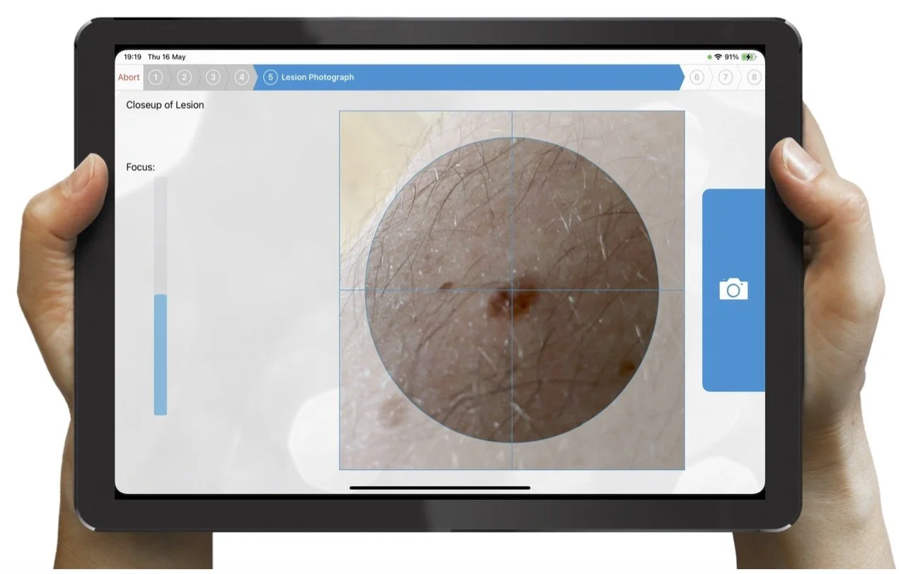
3. Capture lesion
The user takes a close-up of the suspect skin lesion using the iPad camera.

4. Identify edge
The edge is automatically identified and shown green on the image.
The user confirms that the lesion is correctly identified.

5. Result
The result is given immediately as a value representing the chance the lesion is a melanoma together with a graphic to aid interpretation.
6. Report
The Report contains relevant patient information, site and user information, date and time stamp, nomela® reference, nomela® result and images for transmission to ePR.